
The Silent Fragility Crisis: Why Bone Mineral Density (BMD) Is the Overlooked Longevity Biomarker for Denver Athletes & Active Adults
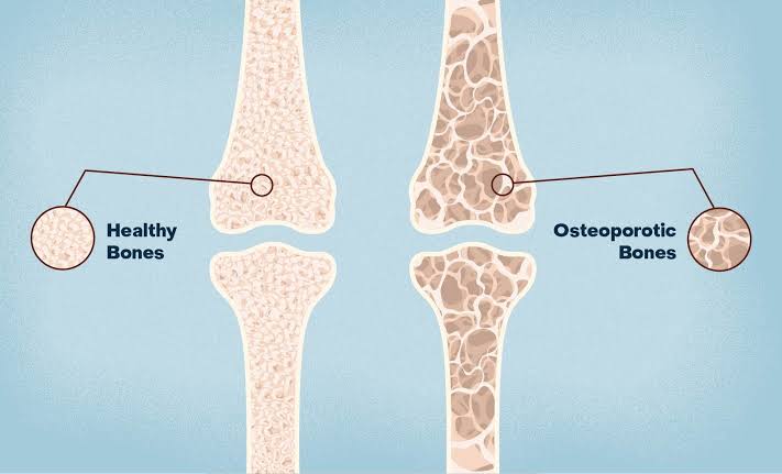
When we talk about healthspan and longevity in Colorado, conversations inevitably gravitate toward high VO2 Max scores, clean ketogenic diets, sub-15% body fat, and rigorous cardiovascular conditioning. Active Mile-High residents spend thousands of hours logging miles along Cherry Creek, crushing mountain bike climbs up Lookout Mountain, and skinning up Berthoud Pass. We measure our resting heart rates with wearables, track our sleep cycles meticulously, and pride ourselves on external fitness.
Yet, deep within our bodies lies a silent, architectural crisis that standard blood panels, fitness watches, and bathroom scales cannot detect: the gradual, asymptomatic decay of our skeletal framework.
Bone Mineral Density (BMD) is arguably the most neglected biomarker in functional longevity medicine today. Medical literature has historically treated bone loss as an inevitable affliction reserved solely for post-menopausal women in their late 70s. However, cutting-edge clinical research—championed by longevity authorities like Dr. Peter Attia—reveals a starkly different reality: skeletal decline begins in our early 30s, progresses silently for decades, and poses one of the single greatest existential threats to long-term mobility, independence, and survival.
Warning — The High-Stakes Reality of Hip Fractures: Clinical epidemiology demonstrates that for adults over the age of 65 who suffer an osteoporotic hip fracture, all-cause mortality within 12 months ranges between 20% and 30%. More critically, over 50% of survivors never regain their pre-fracture functional independence. Skeletal health is not merely about posture—it is a life-or-death metric of longevity.
The Biology of the Living Skeleton: Remodeling, Micro-Architecture & Peak Bone Mass
To understand why bone density demands proactive monitoring, we must dismantle the prevailing myth that bones are inert, static chalksticks. Your skeleton is a dynamic, metabolically vigorous organ that completely turns over and replaces its cellular matrix roughly every 10 years through a tightly orchestrated process known as bone remodeling.
This remodeling cycle hinges on two antagonistic cellular teams:
- Osteoclasts: Specialized multinucleated cells derived from hematopoietic stem cells that secrete hydrochloric acid and cathepsin K (collagenolytic enzymes), dissolving aged, micro-damaged bone tissue (a process called resorption).
- Osteoblasts: Bone-forming cells derived from mesenchymal stem cells that synthesize and deposit new unmineralized organic matrix (osteoid), which subsequently calcifies with hydroxyapatite crystals.
Throughout childhood, adolescence, and our early twenties, osteoblastic synthesis decisively outpaces osteoclastic breakdown. Humans achieve their absolute Peak Bone Mass (PBM) between the ages of 20 and 30. From that milestone forward, hormonal shifts, chronic low-grade inflammation, oxidative stress, and mechanical disuse gradually tip the remodeling scale in favor of resorption.
After age 35, both men and women lose approximately 0.5% to 1.0% of their bone mass every single year. For women traversing the perimenopausal and postmenopausal transition, the abrupt drop in circulating estrogen accelerates this loss up to 2% to 5% per year for a critical 5-to-7-year window.
The Colorado Athlete Paradox: Why High Fitness Does Not Equal Strong Bones
One of the most surprising and sobering patterns we witness at DexaFit Denver is the "fit but fragile" paradox. Mile-High athletes frequently walk into our Auraria Parkway clinic boasting elite VO2 Max scores (often above 55 mL/kg/min) and single-digit visceral fat levels, only to discover their spine or femoral neck T-score sits deep inside the osteopenic range.
How can someone who runs 40 miles a week or bikes 150 miles across the Front Range suffer from premature bone demineralization? The answer lies in biomechanics and energy availability:
- Non-Weight-Bearing Cardiovascular Bias (Cyclists and Swimmers): Bones respond to a phenomenon called mechanotransduction—the biological conversion of mechanical stress and ground-impact shockwaves into biochemical osteoblast-stimulating signals (via fluid shear stress through the canalicular network of osteocytes). While road cycling, gravel biking, and swimming deliver phenomenal cardiovascular adaptations, they are virtually non-weight-bearing. Cyclists sit suspended on a saddle with minimal multi-directional skeletal impact. Without high-magnitude gravitational loading, the body downregulates bone mineral deposition. Multiple sports medicine studies show that competitive master cyclists often possess lower spinal bone density than sedentary non-athletes.
- Relative Energy Deficiency in Sport (RED-S): Denver's endurance culture is exceptionally driven. Trail runners, ultrarunners, and triathletes frequently train in prolonged, unrecognized caloric deficits. When energy intake fails to match exercise expenditure, the body enters a state of low energy availability (LEA). To conserve vital organ function, the hypothalamic-pituitary-gonadal axis suppresses reproductive hormones—reducing circulating estradiol in women and testosterone in men. Both hormones are critical suppressors of osteoclastic activity. In their absence, bones dissolve at an accelerated clip, leading to recurrent stress fractures in metatarsals, tibias, and femoral necks.
- High-Velocity Skiing and Mountain Sports Risk: Colorado's lifestyle is deeply intertwined with winter backcountry skiing, downhill resort runs, and alpine hiking. High-speed collisions, sudden falls on ice, and mogul-impact forces subject the musculoskeletal system to immense torque. An athlete with undiagnosed osteopenia skiing at Vail or Breckenridge faces a drastically elevated risk of catastrophic spiral fractures, tibial plateau breaks, and acetabular trauma from falls that a skeleton with healthy mineral density could easily absorb.
Deconstructing the DEXA Diagnostic Report: T-Scores, Z-Scores & Regions of Interest
Standard screening guidelines in conventional healthcare are reactive rather than preventive. Most insurance policies do not cover bone density scans until a woman turns 65 or a man turns 70—or until after they suffer a traumatic fracture from a ground-level stumble. Waiting until your late 60s to check your bone health is like waiting for a massive myocardial infarction before checking your cholesterol.
Dual-Energy X-ray Absorptiometry (DEXA) is the undisputed medical gold standard for non-invasive, clinical bone mineral assessment. A clinical DEXA scan utilizes two low-dose X-ray beams of distinct energy peaks (high energy and low energy) to distinguish between soft tissue, fat mass, and bone mineral content with sub-millimeter precision.
Note — Radiation Safety Comparison: A full clinical DEXA scan exposes the patient to approximately 1 to 4 microsieverts (µSv) of radiation. For context, a standard dental X-ray delivers ~5 µSv, a single cross-country flight from Denver to New York exposes you to ~20 to 30 µSv of cosmic radiation, and daily natural background radiation in the Mile High City is approximately 8 to 10 µSv. A DEXA scan is extraordinarily safe, gentle, and fast.
When you complete your scan at DexaFit Denver, your clinical report translates raw bone mineral content (in grams per square centimeter, g/cm²) into standardized statistical benchmarks:
Normal Bone Density
- T-Score Range
- -1.0 or higher
- Clinical Meaning
- Within 1 SD of a healthy 30-year-old reference adult.
- Denver Clinical Action Plan
- Maintain progressive resistance training, ensure 1.6g/kg protein, optimize D3/K2. Re-test every 18–24 months.
Osteopenia (Low Bone Mass)
- T-Score Range
- -1.0 to -2.5
- Clinical Meaning
- Early structural demineralization; elevated future fracture risk if untreated.
- Denver Clinical Action Plan
- Implement targeted axial impact loading, evaluate hormone profiles, eliminate RED-S. Re-test annually.
Osteoporosis
- T-Score Range
- -2.5 or lower
- Clinical Meaning
- Severe porous architecture; high risk of fragility and spontaneous fractures.
- Denver Clinical Action Plan
- Medical consultation required; clinical pharmacological intervention, specialized fall-prevention physical therapy.
Severe Osteoporosis
- T-Score Range
- -2.5 or lower + Fracture
- Clinical Meaning
- Compromised micro-architecture accompanied by one or more prior fractures.
- Denver Clinical Action Plan
- Urgent multi-disciplinary endocrinology and orthopedic intervention.
| Classification | T-Score Range | Clinical Meaning | Denver Clinical Action Plan |
|---|---|---|---|
| Normal Bone Density | -1.0 or higher | Within 1 SD of a healthy 30-year-old reference adult. | Maintain progressive resistance training, ensure 1.6g/kg protein, optimize D3/K2. Re-test every 18–24 months. |
| Osteopenia (Low Bone Mass) | -1.0 to -2.5 | Early structural demineralization; elevated future fracture risk if untreated. | Implement targeted axial impact loading, evaluate hormone profiles, eliminate RED-S. Re-test annually. |
| Osteoporosis | -2.5 or lower | Severe porous architecture; high risk of fragility and spontaneous fractures. | Medical consultation required; clinical pharmacological intervention, specialized fall-prevention physical therapy. |
| Severe Osteoporosis | -2.5 or lower + Fracture | Compromised micro-architecture accompanied by one or more prior fractures. | Urgent multi-disciplinary endocrinology and orthopedic intervention. |
Understanding T-Score vs. Z-Score
- T-Score: Compares your bone mineral density to that of a healthy, peak-density 30-year-old adult of your biological sex. This is the primary prognostic score used to diagnose osteopenia and calculate 10-year fracture risk (FRAX).
- Z-Score: Compares your bone mineral density to an age-, sex-, and weight-matched peer group. A Z-score lower than -2.0 indicates secondary causes of accelerated bone loss (such as hyperparathyroidism, celiac malabsorption, prolonged corticosteroid use, or hypogonadism) that require medical investigation.
Key Anatomical Scan Regions
DexaFit's advanced dual-energy system evaluates distinct skeletal regions characterized by different ratios of dense cortical bone and metabolically sensitive trabecular bone:
- Lumbar Spine (L1–L4): Composed predominantly of spongy trabecular bone, which has high metabolic turnover. The lumbar spine is often the earliest site to display rapid demineralization or, conversely, rapid therapeutic recovery from resistance training.
- Dual Femoral Neck and Total Hip: Composed of a mixture of cortical and trabecular architecture. The femoral neck is the single most critical predictor of life-altering hip fractures.
- Forearm (Radius 33%): Frequently evaluated in patients with hyperparathyroidism or when hip/spine artifacts (such as prior hardware or severe lumbar osteoarthritis) prevent accurate regional assessment.
The Deadly Synergistic Threat: Osteosarcopenia
In modern geriatric medicine and sports physiology, clinicians no longer look at bone and muscle in isolation. The emerging clinical paradigm is Osteosarcopenia—the concurrent loss of skeletal muscle mass/strength (sarcopenia) and bone mineral density (osteopenia/osteoporosis).
Muscle and bone form an inseparable mechanical and endocrine unit:
- Mechanical Coupling: Skeletal muscles pull on periosteal attachment points, applying bending moments and torsional forces that stimulate bone calcification.
- Biochemical Crosstalk: Healthy skeletal muscle secretes myokines (such as irisin and IGF-1) that communicate directly with osteoblasts. Conversely, healthy bone secretes osteocalcin, which enhances muscle nutrient uptake and mitochondrial function.
When lean muscle mass deteriorates—often exacerbated by sedentary desk work, crash dieting, or GLP-1 weight loss medications without adequate resistance training—the skeleton loses its primary mechanical stimulus and shock absorber. This leads to a vicious spiral: weaker muscles lead to poorer balance and unsteady gait; decreased bone density ensures that when an inevitable trip or slip occurs on an icy Denver sidewalk, the skeleton shatters instead of flexing.
The Proactive Playbook: How to Rebuild and Preserve Skeletal Density
The prevailing dogma that bone loss is entirely irreversible is scientifically outdated. While reversing late-stage severe osteoporosis requires specialized pharmaceutical bone-forming agents (such as teriparatide or romosozumab), individuals with normal density or early-stage osteopenia can actively stabilize and even increase bone mineral density through targeted physiological stimuli:
High-Intensity Resistance Training (Axial Loading): To trigger mechanotransduction, the skeletal system requires mechanical strain that exceeds ordinary daily thresholds. The landmark LIFTMOR clinical trials published in the Journal of Bone and Mineral Research demonstrated that high-intensity, low-repetition resistance training (80–85% 1-Rep Max) safely increases bone mineral density in both the lumbar spine and femoral neck of individuals with low bone mass.
The most effective multi-joint compound exercises include:
- Barbell Back Squats & Front Squats: Transmits compressive axial force down the entire spinal column, sacrum, and femur.
- Conventional Deadlifts & Trap-Bar Deadlifts: Provides massive tensile load through the hips, pelvic girdle, and upper posterior chain.
- Overhead Presses & Push Presses: Stresses the clavicles, humerus, and thoracic vertebrae.
- Weighted Carries (Farmer's Walks): Imparts vertical gravitational loading while reinforcing grip and hip stabilizer recruitment.
Multi-Directional Dynamic Impact Loading: Unlike steady-state cycling or rowing, dynamic impact loading delivers fast strain rates that "wake up" dormant osteocytes. Incorporating 50 to 100 high-impact ground strikes per session—such as jump roping, plyometric box jumps, bounding, tennis, and trail running downhill—provides the diverse impact vectors needed to stimulate femoral and calcaneal bone remodeling.
Micronutrient Synergy: Beyond Calcium Carbonate
Chalky calcium supplements taken in isolation are ineffective and can contribute to arterial calcification. Bone mineralization requires an orchestrating symphony of co-factors:
- Elemental Calcium (1,000–1,200 mg/day): Best obtained from whole food sources such as fermented dairy (kefir, Greek yogurt), canned wild salmon with bones, sardines, and leafy greens.
- Vitamin D3 (2,000–5,000 IU/day): Facilitates intestinal calcium absorption. (Given Denver's winter altitude, serum 25-hydroxyvitamin D levels often plummet between November and April).
- Vitamin K2 (MK-7, 100–200 mcg/day): Activates osteocalcin, the molecular glue that anchors calcium ions directly into the hydroxyapatite bone matrix while preventing calcium from depositing in arterial walls.
- Magnesium Glycinate or Malate (300–400 mg/day): Essential co-factor required for enzymatic conversion of vitamin D into its active hormonal form (calcitriol).
- Dietary Protein (1.6 to 2.2 g/kg/day): Approximately 50% of bone volume is composed of protein (primarily Type 1 collagen fibers). Inadequate dietary protein triggers secondary hyperparathyroidism, stripping minerals from the skeleton.
What to Expect During Your DEXA Bone Mineral Scan at DexaFit Denver
Visiting DexaFit Denver for a comprehensive clinical DEXA scan is an efficient, relaxing, and deeply informative experience designed to give you clarity and complete ownership over your internal health metrics.
- 1. Pre-Test Preparation: You arrive at our clean, state-of-the-art Denver clinic wearing comfortable athletic clothing free of metal zippers, snaps, underwires, or metallic belts. You should refrain from taking high-dose calcium supplements for 24 hours prior to testing.
- 2. Patient Positioning: You lie completely flat on your back on a cushioned scanning table. Our certified technicians position your body with precision, using specialized positioning blocks to align your hips and lumbar spine for clinical reproducibility.
- 3. The Scan: The mechanical scanner arm passes smoothly and silently over your body from head to toe over the course of 6 to 10 minutes. The scan is completely painless, open-air, non-claustrophobic, and whisper-quiet.
- 4. Immediate Results & Data Review: Immediately upon completion, you receive a full-color, medical-grade report detailing your regional bone mineral density, T-scores, Z-scores, visceral fat volume, and bilateral lean muscle mass symmetry. Our clinical team walks you through every metric to ensure you understand your baseline.
This article is for educational purposes and is not medical advice, diagnosis, or treatment. Consult a qualified healthcare provider before starting supplements, changing medications, or beginning a new high-intensity training program, especially if you have been diagnosed with osteopenia or osteoporosis.
References
- Watson, S. L., et al. (2018). High-Intensity Resistance and Impact Training Improves Bone Mineral Density and Physical Function in Postmenopausal Women with Osteopenia and Osteoporosis: The LIFTMOR Trial. Journal of Bone and Mineral Research, 33(2), 211–220.
- Attia, P., & Gifford, B. (2023). Outlive: The Science and Art of Longevity. Harmony Books/Crown Publishing, New York.
- Borer, K. T. (2005). Physical activity in the prevention and amelioration of osteoporosis in women: interaction of mechanical, hormonal and dietary factors. Sports Medicine, 35(9), 779–830.
- Mountjoy, M., et al. (2018). The IOC consensus statement: relative energy deficiency in sport (RED-S). British Journal of Sports Medicine, 52(11), 687–697.
- World Health Organization (WHO). (2004). Assessment of fracture risk and its application to screening for postmenopausal osteoporosis. WHO Technical Report Series.
- Scofield, K. L., & Hecht, S. (2012). Bone health in endurance athletes: runners, cyclists, and swimmers. Current Sports Medicine Reports, 11(6), 328–334.
Know Your Bone Density Before a Fracture Does
Get regional bone mineral density, T-scores, Z-scores, visceral fat, and lean muscle mass in one medical-grade DEXA report at DexaFit Denver.
Book Your DEXA Bone Density Scan